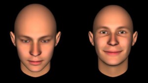
Uno studio che ha osservato i deficit di pazienti neurolesi dimostra che nell’ imitazione di gesti sono coinvolte almeno due componenti elaborate ognuna da un emisfero diverso.
SISSA, Scuola Internazionale Superiore di Studi Avanzati
Imitando si imparano un sacco di cose: a camminare, a suonare uno strumento, fare uno sport e tanto altro ancora. Quali sono i processi nel cervello alla base dell’imitazione? Da qualche anno ormai la scienza ha scoperto il ruolo dei neuroni specchio, ma ancora molto resta da capire. Uno studio che ha osservato i deficit di pazienti neurolesi dimostra che nell’imitazione di gesti sono coinvolte almeno due componenti elaborate ognuna da un emisfero diverso. Lo studio cui ha partecipato la SISSA è stato pubblicato su Neuropsychologia.
Imitazione di gesti dopo una lesione cerebrale: aprassia ideomotoria
Dopo una lesione cerebrale (per esempio causata da ictus o emorragia) si può sviluppare una difficoltà selettiva nell’imitare i gesti e i movimenti altrui (aprassia ideomotoria). Nella storia della neuropsicologia questi studi sono fra i più noti (i primi risalgono addirittura all’inizio ‘900), anche perché questi deficit ostacolano gli interventi terapeutici volti al recupero di abilità motorie, visto che il paziente non può eseguire, imitandoli, i gesti del medico. Negli ultimi vent’anni questi studi hanno poi trovato nuovo vigore grazie alla scoperta dei neuroni specchio, eppure si sa ancora troppo poco di questi processi. Molti scienziati pensano che vi sia un ruolo determinante dell’emisfero sinistro, visto che nella maggior parte sono proprio i cerebrolesi unilaterali sinistri a mostrare questo disturbo. Ma come spiegare allora anche una piccola percentuale di pazienti aprassici con una lesione unilaterale destra?
Paola Mengotti, alla SISSA al tempo dello studio, ora al Forschungszentrum di Jülich in Germania, Raffaella Rumiati, professoressa della SISSA e responsabile del laboratorio iNSuLa (Neuroscience and Society), e colleghi hanno condotto uno studio per rispondere a questa domanda.
Alla ricerca hanno partecipato venti pazienti (che sono stati visti all’Ospedale San Camillo di Venezia e all’Azienda Ospedaliero-Universitaria Ospedali Riuniti di Trieste) con lesioni unilaterali sinistre o destre, più un gruppo di controllo. L’idea di partenza era che l’imitazione fosse composta almeno da due compiti distinti: quello dell’imitazione motoria e un compito spaziale. Quando infatti dobbiamo imitare i movimenti di qualcuno, non solo ripetiamo i suoi gesti, ma dobbiamo anche traslarli sul nostro corpo (in pratica dobbiamo rispecchiarli). Nello studio i pazienti eseguivano compiti di imitazione in una delle due componenti, imitazione motoria e imitazione spaziale. Sono poi state confrontate le prestazioni per ogni componente e messe in relazione al tipo di lesione.
Quel che è emerso è che nella performance imitativa conta la somiglianza fra quanto visto e quanto prodotto, e questo ovviamente interagisce con il tipo di deficit dell’individuo.
[blockquote style=”1″] Analizzando le prestazioni dei pazienti con lesioni dell’emisfero destro e dell’emisfero sinistro con due compiti di imitazione, abbiamo potuto dimostrare che l’imitazione si basa sulla somiglianza tra l’azione osservata e quella prodotta[/blockquote]
spiega Rumiati.
[blockquote style=”1″]Questa somiglianza riflette o una corrispondenza anatomica o una spaziale. Lesioni dell’emisfero sinistro compromettono la prima operazione mentre lesioni dell’emisfero destro compromettono la seconda.[/blockquote]
LINK UTILI: • Articolo originale su Neuropsychologia: http://goo.gl/DjV6QG
IMMAGINI: • Crediti: Jamie McCaffrey (Flickr: https://goo.gl/5XffDR)
Contatti: Ufficio stampa: [email protected] Tel: (+39) 040 3787644 | (+39) 366-3677586
via Bonomea, 265, 34136 Trieste
Maggiori informazioni sulla SISSA
One study focusing on neurological patients showed that at least two components are involved in imitating gestures, each from a different hemisphere of the brain.
We learn many things through imitation: how to walk, play an instument, sports, and even more. What are the processes in the brain responsible for imitation? For some years now, science has been examining the role of mirror neurons, but there is still much to understand. One study focusing on neurological patients showed that at least two components are involved in imitating gestures, each from a different hemisphere of the brain. The study, which SISSA participated in, was published in Neuropsychologia.
After a brain injury (caused by stroke or hemorrhage, for example), patients may have difficulty imitating gestures and movements of others (ideomotor apraxia). In the history of neuropsychology, these studies are among the best known (the first date back to the early 1900’s) as these deficits hinder therapy aimed at recovering motor skills, since the patient cannot perform gestures by imitating the doctor. In the last twenty years, these studies have found new significance thanks to the discovery of mirror neurons, and yet little is known about these processes. Many scientists think the left hemisphere plays a dominant role because this problem most often surfaces in cases of unilateral brain-damage of the left hemisphere. How then, can we explain the small percentage of apraxic patients who have suffered unilateral lesions to the right hemisphere?
Paola Mengotti, at SISSA at the time of the study, now at Forschungszentrum Jülich in Germany, SISSA Professor and Head of the iNSuLa Laboratory (Neuroscience and Society), Raffaella Rumiati, and colleagues conducted a study to answer this question. Twenty patients (visited at San Camillo in Venice and Azienda Ospedaliero-Universitaria Ospedali Riuniti in Trieste) with unilateral brain lesions in the left or right hemispheres, plus a control group participated in the study. The initial idea was that imitation is made up of at least two distinct tasks: motor imitation, and a spatial component. When we have to imitate someone else’s movements, we not only have to repeat the actions, but we also have to translate them to our body (mirror them). In the study, patients performed imitation tasks using one of the two components, motor or spatial. Performance for each component was then compared and categorized in relation to the type of lesion.
What emerged is that what counts in imitation is the similarity between what is seen and what is produced, and this of course depends on the individual type of deficit. [blockquote style=”1″]Analyzing the performance of two imitation tasks by patients with lesions in the right hemisphere and the left hemisphere, we were able to demonstrate that imitation is based on the similarity between the observed action and the one produced[/blockquote] explains Rumiati. [blockquote style=”1″]This similarity reflects either an anatomical match or a spatial one. Lesions in the left hemisphere affect the former while lesions in the right hemisphere affect the latter.[/blockquote]
USEFUL LINKS: • Original paper on Neuropsychologia: http://goo.gl/DjV6QG
IMAGE: • Credits: Jamie McCaffrey (Flickr: https://goo.gl/5XffDR)
Contacts: Press office: [email protected] Tel: (+39) 040 3787644 | (+39) 366-3677586
via Bonomea, 265 34136 Trieste


